
Deuteranopia – Red-Green Color Blindness
Deutan color vision deficiencies are by far the most common forms of color blindness. This subtype of red-green color blindness is found in about 6% of the male population, mostly in its mild form deuteranomaly.
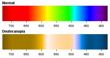
Normal and Deuteranopia Color Spectrum
When you have a look at the color spectrum of a deuteranopic person you can see that a variety of colors look different than in a normal color spectrum. Whereas red and green are the main problem colors, there are also for example some gray, purple and a greenish blue-green which can’t be distinguished very well.
The well known term red-green color blindness is actually split into two different subtypes. On one side persons which either lack or have anomalous long wavelength sensitive cones (protan color vision deficiency), which are more responsible for the red part of vision. And on the other side deutan color vision deficiencies, which again are split into two different types:
- Dichromats: Deuteranopia (also called green-blind). In this case the medium wavelength sensitive cones (green) are missing at all. A deuteranope can only distinguish 2 to 3 different hues, whereas somebody with normal vision sees 7 different hues.
- Anomalous Trichromats: Deuteranomaly (green-weak). This can be everything between almost normal color vision and deuteranopia. The green sensitive cones are not missing in this case, but the peak of sensitivity is moved towards the red sensitive cones.
Below you can see a picture with normal colors on the left side and altered colors on the right side. The picture on the right side shows you how a person affected by deuteranopia would see the scenery (picture taken by Ottmar Liebert, some rights reserverd).
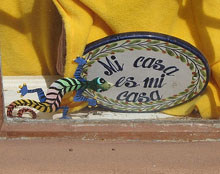 |  |
| Normal Vision | Deuteranopic Vision |
In the midst of the last century there were different researches published concerning unilateral deuteranopia. Some persons were found which had trichromatic vision in one eye and dichromatic vision in the other. The eye with dichromatic vision had a color specturm related to a deuteranopia color spectrum. One case of such a one-eyed colorblind is described in the article The Spectral Luminosity Curves for a Dichromatic Eye and a Normal Eye in the Same Person.
The one-eyed color blindness is definitely not the common case, whereas deuteranopia and especially deuteranomaly are the most observed cases of all color vision deficiencies. In 75% of all occurrences of color blindness it is a defect caused by the green sensitive cones. The following list shows the approximative rates of deutan defects in our population:
- Deuteranomaly, Male Population: 5%
- Deuteranopia, Male Population: 1%
- Deuteranomaly, Female Population: 0.35%
- Deuteranopia, Female Population: 0.1%
These numbers don’t change much, because deutan color blindness as one form of red-green color blindness is a congenital disease. Red-green color blindness is a sex-linked trait and therefore encoded on the X chromosome. Because women have two X and can overcome the handicap of one, men have only one and are therefore more often affected. This circumstance can also be read in the numbers of the table above. More details about the concrete inheritance pattern can be found at The Biology behind Red-Green Color Blindness.
If you are colorblind there is a big chance that you are red-green colorblind, usually green-weak and male. And specially if you are suffering from deuteranomaly, this condition is not as rare as you might think and you even might find some of your friends who’s also suffering under this color vision deficiency.
Read more about Tritanopia and Protanopia—the other two types of color blindness.
The Biology behind Red-Green Color Blindness
Red-green color blindness is in the majority of cases provoked through a defective X-chromosome. Human beings have 23 different pairs of chromosomes whereof one pair is the so called sex-chromosome. This pair consists of two X-chromosomes on women and one X- coupled with one Y-chromosome on men. Color vision in the red-green area is coded on the X-chromosome which is called a sex linked trait.
This concludes if a man is a carrier of a defective X-chromosome he will suffer from color blindness. On women the not defective chromosome is in charge and therefore she is not colorblind but a carrier for color blindness. Because a women needs two defective X-chromosome to be affected this symptome is called X-linked recessive. A very interesting conclusion of this: If you are male and your father suffers from a red-green color vision deficiency you can not inherit it from him. Only women can be carriers for color blindness who pass it on to their sons.
 | 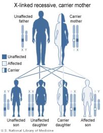 |
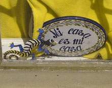
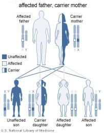
Let’s have a look at some illustrations. On the left you can see how the disorder is passed on from an affected father to his children. The sons are unaffected and do not have the mutation. The daughters are not affected but are both carriers of the disorder because they inherited the defective X-chromosome from their father. The illustration on the right side shows a mother which is a carrier and a father which is unaffected. Their son is at a rate of 50% affected i.e. red-green colorblind and their daughter is at the same rate either are carrier or unaffected.
X link Recessive Mother And Father
In the last illustration we coupled an affected man with a women which is a carrier. As you can see their children are at a rate of 50% affected. This is the only case shown here, where a women can be affected i.e. suffering from a red-green color blindness. If the children are unaffected the daughter is anyway a carrier of the disorder. The not shown combinations where man and women are either both affected or both unaffected are left to the reader…
I hope this could give a better insight into the biology behind color blindness. It has to be noted that these remarks are only true for red-green color blindness. Blue-yellow color blindness (tritanopia) is linked to the chromosome pair 7 and therefore sex independent. Further readings on this topic and more details can be found under the following links:
The above illustrations are provided by the U.S. National Library of Medicine, 7th March 2006.
https://www.color-blindness.com/2006/03/07/the-biology-behind/
No comments:
Post a Comment