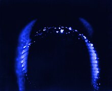
Kirlian photography
From Wikipedia, the free encyclopedia
Kirlian photography is a collection of photographic techniques used to capture the phenomenon of electrical coronal discharges. It is named after Semyon Kirlian, who, in 1939 accidentally discovered that if an object on a photographic plate is connected to a high-voltage source, an image is produced on the photographic plate.[1] The technique has been variously known as "electrography",[2] "electrophotography",[3] "corona discharge photography" (CDP),[4] "bioelectrography",[2] "gas discharge visualization (GDV)",[5] "eletrophotonic imaging (EPI)",[6] and, in Russian literature, "Kirlianography".
Kirlian photography has been the subject of mainstream scientific research, parapsychology research and art. To a large extent, It has been co-opted by promoters of pseudoscience and paranormal health claims in books, magazines, workshops, and web sites.[7][8]
History
In 1889, Czech B. Navratil coined the word "electrography". Seven years later in 1896, a French experimenter, H. Baravuc, created electrographs of hands and leaves.
In 1898, Russian engineer Yakov Narkevich-Iodko[9][note 1] demonstrated electrography at the fifth exhibition of the Russian Technical Society.
In 1939, two Czechs, S. Pratt and J. Schlemmer published photographs showing a glow around leaves. The same year, Russian electrical engineer Semyon Kirlian and his wife Valentina developed Kirlian photography after observing a patient in Krasnodar hospital who was receiving medical treatment from a high-frequency electrical generator. They had noticed that when the electrodes were brought near the patient's skin, there was a glow similar to that of a neon discharge tube.[10]
The Kirlians conducted experiments in which photographic film was placed on top of a conducting plate, and another conductor was attached to the a hand, a leaf or other plant material. The conductors were energized by a high frequency high voltage power source, producing photographic images typically showing a silhouette of the object surrounded by an aura of light.
In 1958, the Kirlians reported the results of their experiments for the first time. Their work was virtually unknown until 1970, when two Americans, Lynn Schroeder and Sheila Ostrander published a book, Psychic Discoveries Behind the Iron Curtain. Although little interest was generated among western scientists, Russians held a conference on the subject in 1972, at Kazakh State University.[11]
Kirlian photography was used extensively in the former Eastern Bloc. For example, in the 1970s, Romania had 14,000 state-sponsored scientists working on the technique.[12] The corona discharge glow at the surface of an object subjected to a high voltage electrical field is referred to as a Kirlian aura in Russia and Eastern Europe,[13][14] however this should not to be confused with the paranormal concept of the aura. In 1975 Belarusian scientist Victor Adamenko wrote a dissertation titled Research of the structure of High-frequency electric discharge (Kirlain effect) images.[15][16][17][18]
Early in the 1970s, Thelma Moss and Kendall Johnson at the Center for Health Sciences at the UCLA conducted extensive research[11] into Kirlian photography. Moss led an independent and unsupported parapsychology laboratory[19] that was shut down by the university in 1979.[20]
Kirlian's research first became known in the United States after Shelia Ostrander's and Lynn Schroeder's book "Psychic Discoveries Behind the Iron Curtain" was published in 1970. High voltage electrophotography soon became known to the general public as Kirlian Photography.
Overview
Kirlian photography is a technique for creating contact print photographs using high voltage. The process entails placing sheet photographic film on top of a metal discharge plate. The object to be photographed is then placed directly on top of the film. High voltage is momentarily applied to the metal plate, thus creating an exposure. The corona discharge between the object and the high voltage plate is captured by the film. The developed film results in a Kirlian photograph of the object.
Color photographic film is calibrated to faithfully produce colors when exposed to normal light. Corona discharges can interact with minute variations in the different layers of dye used in the film, resulting in a wide variety of colors depending on the local intensity of the discharge.[21] Film and digital imaging techniques also record light produced by photons emitted during corona discharge (see Mechanism of corona discharge).
Photographs of inanimate objects such as a coins, keys and leaves can be made more effectively by grounding the object to the earth, a cold water pipe or to the opposite (polarity) side of the high voltage source. Grounding the object creates a stronger corona discharge.[22]
Kirlian photography does not require the use of a camera or a lens because it is a contact print process. It is possible to use a transparent electrode in place of the high voltage discharge plate, allowing one to capture the resulting corona discharge with a standard camera or a video camera.[23]
Visual artists such as Robert Buelteman,[24] Ted Hiebert,[25] and Dick Lane[26] have used Kirlian photography to produce artistic images of a variety of subjects. Kirlian Photographer Mark D. Roberts, who has worked with Kirlian imagery for over 40 years, published a portfolio of plant images entitled "Vita occulta plantarum" or "The Secret Life of Plants", first exhibited in 2012 at the Bakken Museum in Minneapolis.
In 1968, Antonov photographed water droplets using black and white Kirlian photography.[32] Similar research was carried out by Ignat Ignatov almost 40 years later.
In 2010, Ignatov used color Kirlian photography to study droplets of different types of water.[37] He found that his results depend on the dielectric permittivity of water, which is related to the composition and spectral analysis of water. Kirlian photography is used as an auxiliary method for Bulgarian scientists in research the properties of water. The primary method used is spectral analysis in the infrared spectrum.[38][39][40]
Galina Gudakova conducted biological research in Russia.[46][47] She explored the growth of microbiological cultures using Kirlian photograph.
The living aura theory is at least partially repudiated by demonstrating that leaf moisture content has a pronounced effect on the electric discharge coronas; more moisture creates larger, more dynamic corona discharges. As the leaf dehydrates, the coronas will naturally decrease in variability and intensity. As a result, the changing water content of the leaf can affect the so-called Kirlian aura. Kirlian's experiments did not provide evidence for an energy field other than the electric fields produced by chemical processes, and the streaming process of coronal discharges.[4]
The coronal discharges identified as Kirlian auras are the result of stochastic electric ionization processes, and are greatly affected by many factors, including the voltage and frequency of the stimulus, the pressure with which a person or object touches the imaging surface, the local humidity around the object being imaged, how well grounded the person or object is, and other local factors affecting the conductivity of the person or object being imaged. Oils, sweat, bacteria, and other ionizing contaminants found on living tissues can also affect the resulting images.[51][52][53]
http://en.wikipedia.org/wiki/Kirlian_photography
Kirlian photography has been the subject of mainstream scientific research, parapsychology research and art. To a large extent, It has been co-opted by promoters of pseudoscience and paranormal health claims in books, magazines, workshops, and web sites.[7][8]
History
In 1889, Czech B. Navratil coined the word "electrography". Seven years later in 1896, a French experimenter, H. Baravuc, created electrographs of hands and leaves.
In 1898, Russian engineer Yakov Narkevich-Iodko[9][note 1] demonstrated electrography at the fifth exhibition of the Russian Technical Society.
In 1939, two Czechs, S. Pratt and J. Schlemmer published photographs showing a glow around leaves. The same year, Russian electrical engineer Semyon Kirlian and his wife Valentina developed Kirlian photography after observing a patient in Krasnodar hospital who was receiving medical treatment from a high-frequency electrical generator. They had noticed that when the electrodes were brought near the patient's skin, there was a glow similar to that of a neon discharge tube.[10]
The Kirlians conducted experiments in which photographic film was placed on top of a conducting plate, and another conductor was attached to the a hand, a leaf or other plant material. The conductors were energized by a high frequency high voltage power source, producing photographic images typically showing a silhouette of the object surrounded by an aura of light.
In 1958, the Kirlians reported the results of their experiments for the first time. Their work was virtually unknown until 1970, when two Americans, Lynn Schroeder and Sheila Ostrander published a book, Psychic Discoveries Behind the Iron Curtain. Although little interest was generated among western scientists, Russians held a conference on the subject in 1972, at Kazakh State University.[11]
Kirlian photography was used extensively in the former Eastern Bloc. For example, in the 1970s, Romania had 14,000 state-sponsored scientists working on the technique.[12] The corona discharge glow at the surface of an object subjected to a high voltage electrical field is referred to as a Kirlian aura in Russia and Eastern Europe,[13][14] however this should not to be confused with the paranormal concept of the aura. In 1975 Belarusian scientist Victor Adamenko wrote a dissertation titled Research of the structure of High-frequency electric discharge (Kirlain effect) images.[15][16][17][18]
Early in the 1970s, Thelma Moss and Kendall Johnson at the Center for Health Sciences at the UCLA conducted extensive research[11] into Kirlian photography. Moss led an independent and unsupported parapsychology laboratory[19] that was shut down by the university in 1979.[20]
Kirlian's research first became known in the United States after Shelia Ostrander's and Lynn Schroeder's book "Psychic Discoveries Behind the Iron Curtain" was published in 1970. High voltage electrophotography soon became known to the general public as Kirlian Photography.
Overview
| Typical Kirlian photography setup (cross section) |
Color photographic film is calibrated to faithfully produce colors when exposed to normal light. Corona discharges can interact with minute variations in the different layers of dye used in the film, resulting in a wide variety of colors depending on the local intensity of the discharge.[21] Film and digital imaging techniques also record light produced by photons emitted during corona discharge (see Mechanism of corona discharge).
Photographs of inanimate objects such as a coins, keys and leaves can be made more effectively by grounding the object to the earth, a cold water pipe or to the opposite (polarity) side of the high voltage source. Grounding the object creates a stronger corona discharge.[22]
Kirlian photography does not require the use of a camera or a lens because it is a contact print process. It is possible to use a transparent electrode in place of the high voltage discharge plate, allowing one to capture the resulting corona discharge with a standard camera or a video camera.[23]
Visual artists such as Robert Buelteman,[24] Ted Hiebert,[25] and Dick Lane[26] have used Kirlian photography to produce artistic images of a variety of subjects. Kirlian Photographer Mark D. Roberts, who has worked with Kirlian imagery for over 40 years, published a portfolio of plant images entitled "Vita occulta plantarum" or "The Secret Life of Plants", first exhibited in 2012 at the Bakken Museum in Minneapolis.
[edit] Research
Kirlian photography has been a subject of scientific research, parapsychology research and pseudoscientific claims.[7][8] There are no clear delineations between classic scientific research, fringe research, and claims made by promoters of alternative medicine and the like. Much of the research conducted around the middle of the 20th century occurred in the former Eastern Bloc before the cold war ended and has not held up to the scrutiny of stricter Western scientific standards.[edit] Scientific research
Results of scientific experiments published in 1976 involving Kirlian photography of living tissue (human finger tips) showed that most of the variations in corona discharge streamer length, density, curvature and color can be accounted for by the moisture content on the surface of and within the living tissue.[27] Scientists outside of the US have also conducted scientific research.[edit] Anton Antonov
Bulgarian scientist Anton Antonov conducted experiments which show that the conductivity of an object does not influence the resulting images, but that corona discharge formation depends on the distribution of dielectric permittivity. In 1979, Antonov's Kirlian photography experiments generated data about the distribution of the electric field in the air gap between an object and the registering medium during corona discharge. These discharges were found to be formed by both negative nitrogen ions and positive oxygen ions.[28][29][30][31]In 1968, Antonov photographed water droplets using black and white Kirlian photography.[32] Similar research was carried out by Ignat Ignatov almost 40 years later.
[edit] Ignat Ignatov
Ignat Ignatov conducted Kirlian photography research using color film to examine the colors of corona discharges. His experiments showed a correspondence between electron energy levels and the colors recorded on the film. He demonstrated that red corresponds to an energy level of 1.82 electron volts (еV); orange, 2.05 eV; yellow, 2.14 eV; blue-green (cyan), 2.43 eV; blue, 2.64 eV; and violet, 3.03 eV. Green discharge emissions were not observed in these experiments. Ignatov found that image quality on film was much higher than when corona discharges were recorded with digital imaging techniques.[33][34][35][36]In 2010, Ignatov used color Kirlian photography to study droplets of different types of water.[37] He found that his results depend on the dielectric permittivity of water, which is related to the composition and spectral analysis of water. Kirlian photography is used as an auxiliary method for Bulgarian scientists in research the properties of water. The primary method used is spectral analysis in the infrared spectrum.[38][39][40]
[edit] Konstantin Korotkov
Konstantin Korotkov developed a technique similar to Kirlian photography called Gas Discharge Visualization (GDV).[41][42][43] Korotkov's GDV camera system consists of hardware and software to directly record, process and interpret GDV images with a computer. Although diagnostic medicine experiments conducted by Korotkov using GDV were deemed to be statistically unreliable, his web site still promotes his device and research in a medical context.[44][45][edit] Other scientific research
Izabela Ciesielska at the Institute of Architecture of Textiles in Poland experimented with corona discharge photography (CDP) to evaluate the effects of human contact with various textiles on biological factors such as heart rate and blood pressure, as well as corona discharge images. The experiments used the The GDV camera designed by Konstantin Korotkov to capture corona discharge images of subjects fingertips while the subjects wore sleeves of various natural and synthetic materials on their forearms. The results failed to establish a relationship between human contact with the textiles and the corona discharge images, and were considered inconclusive.[9]Galina Gudakova conducted biological research in Russia.[46][47] She explored the growth of microbiological cultures using Kirlian photograph.
[edit] Parapsychology research
Around the 1970s, interest in paranormal research peaked. In 1968, Dr. Thelma Moss, a psychology professor headed UCLA’s Neuropsychiatric Institute (NPI ), which was later renamed the Semel Institute. The NPI had a laboratory dedicated to parapsychology research and staffed mostly with volunteers. The lab was unfunded, unsanctioned and eventually shut down by the university. Toward the end of her tenure at UCLA, Moss became interested in Kirlian photography, a technique that supposedly measured the “auras” of a living being. According to Kerry Gaynor, one of her former research assistants, "many felt Kirlian photography’s effects were just a natural occurrence."[20][edit] Pseudoscientific claims
Kirlian believed that images created by Kirlian photography might depict a conjectural energy field, or aura, thought, by some, to surround living things. Kirlian and his wife were convinced that their images showed a life force or energy field that reflected the physical and emotional states of their living subjects. They thought these images could be used to diagnose illnesses. In 1961, they published their first paper on the subject in the Russian Journal of Scientific and Applied Photography.[48] Kirlian's claims were embraced by energy treatments practitioners.[49][edit] Torn leaf experiment
A typical demonstration used as evidence for the existence of these energy fields involved taking Kirlian photographs of a picked leaf at set intervals. The gradual withering of the leaf was thought to correspond with a decline in the strength of the aura. In some experiments, if a section of a leaf was torn away after the first photograph, a faint image of the missing section would sometimes remain when a second photograph was taken. If the imaging surface is cleaned of contaminants and residual moisture before the second image is taken, then no image of the missing section would appear.[50]The living aura theory is at least partially repudiated by demonstrating that leaf moisture content has a pronounced effect on the electric discharge coronas; more moisture creates larger, more dynamic corona discharges. As the leaf dehydrates, the coronas will naturally decrease in variability and intensity. As a result, the changing water content of the leaf can affect the so-called Kirlian aura. Kirlian's experiments did not provide evidence for an energy field other than the electric fields produced by chemical processes, and the streaming process of coronal discharges.[4]
The coronal discharges identified as Kirlian auras are the result of stochastic electric ionization processes, and are greatly affected by many factors, including the voltage and frequency of the stimulus, the pressure with which a person or object touches the imaging surface, the local humidity around the object being imaged, how well grounded the person or object is, and other local factors affecting the conductivity of the person or object being imaged. Oils, sweat, bacteria, and other ionizing contaminants found on living tissues can also affect the resulting images.[51][52][53]
[edit] Qi
Scientists such as Beverly Rubik have explored the idea of a human biofield using Kirlian photography research, attempting to explain the Chinese discipline of Qigong. Qigong teaches that there is a vitalistic energy called qi (or chi) that permeates all living things. The existence of qi has been mostly rejected by the scientific community. Rubik's experiments relied on Konstantin Korotkov's GDV device to produce images which were thought to visualize these qi biofields in chronically ill patients. Rubik acknowledges that the small sample size in her experiments "was too small to permit a meaningful statistical analysis."[54] Vitalistic energies, such as qi, if they exist, would exist beyond the natural world. Claims that these energies can be captured by special photographic equipment are criticized by skeptics.[49]http://en.wikipedia.org/wiki/Kirlian_photography


No comments:
Post a Comment